
It is when cancer spreads from a tumour to elsewhere in the body that it most often proves fatal. This process of individual cells breaking off from one tumour and moving elsewhere in the body through the circulatory system is the first step in “metastasis” which is the spread of cancer from one organ to another.
New research at the University of Alberta for the first time provides video evidence of how cancer cells move through blood vessel walls and actually find their way into other body tissues.
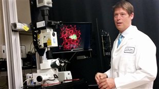
John Lewis (PhD) supervised the research. He is an associate professor in the Department of Oncology and the Sojonky Chair in Prostate Cancer Research. Listen
The research was published in the online science journal Cell Reports in the August 28 edition.
The research team included members from the Lawson Health Research Institute of London Ontario, and the Sandford Burnham Institute in California.
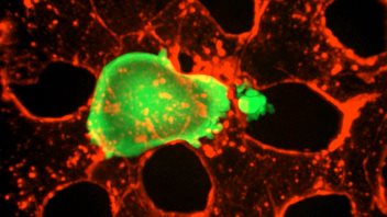
Professor Lewis says it’s been known that cancer cells can produce little extensions, tentacle or fingers if you will, but it was not known until now that they were used to break through blood vessel walls into other parts of the body.
Through their advanced microscopy research Lewis and his team were able to film this process for the first time in a live model.
Their surprising finding is that when a cancer cell gets trapped in a microscopic capillary in the body, it moves around a bit, but then stops and develops tentacles called “invadopodia” to seek out the spaces between cells.
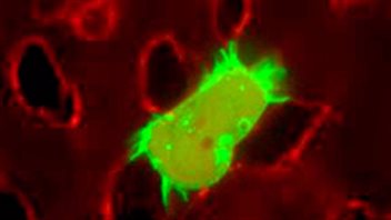
Through chemical activity using enzymes, the invadopodium tentacle breaks down some of the cell material to widen the gap and then the cancer cell follows the tentacle through into the body tissue.
The process is known as “extravasation”.
Once established outside the blood vessel it can divide, mutate, and grow into a new tumour.
Lewis and his team’s research also showed that through drugs or other methods you can inhibit the growth of the tentacles, and so the cancer cell cannot get into the tissue.
He notes that there are limitations to this discovery however.
He says by the time many cancers are detected, metastasis has already taken place. However, pharmaceutical disruption of invadopodia development could be important during surgical procedures.
He says there is evidence that during biopsies or removal of tumours, some cells are broken off and circulate around the body leading to metastasis sometime later.
He says this research could lead to treatment to prevent metastasis during such procedures. He also notes that understanding extravasation is an important aspect in the fight against cancer, and that this research could very well prove to be a big step forward in eventually preventing metastasis and extravasion.
This study was funded by the Canadian Cancer Society Research Institute, Prostate Cancer Canada, Alberta Cancer Foundation, NSERC, and Canadian Breast Cancer Foundation


For reasons beyond our control, and for an undetermined period of time, our comment section is now closed. However, our social networks remain open to your contributions.